Figure 16. Core Infarct (Matched Defect: LOW CBF/LOW CBV)
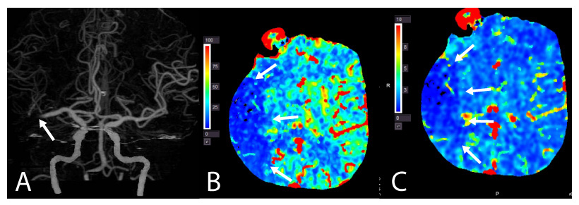
A 68-year–old woman presented with left hemiparesis, dysarthria and neglect. National Institutes of Health Stroke Scale (NIHSS) 19. (A) CT angiography revealed a distal right M1 MCA occlusion. CT perfusion demonstrated (B) reduced right hemispheric cerebral blood flow (CBF) and (C) cerebral blood volume (CBV) indicating a matched defect and thus, core infarct.
Figure 17. Ischemic Penumbra (Mismatched Defect: LOW CBF/HIGH CBV)
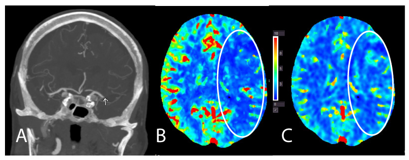
A 96-year–old woman presented with right hemiparesis and global aphasia. National Institutes of Health Stroke Scale (NIHSS) 24. (A) CT angiography revealed a distal left M1 MCA occlusion. CT perfusion demonstrated (B) reduced left hemispheric cerebral blood flow (CBF) and (C) maintained cerebral blood volume (CBV) indicating a mismatch and thus, salvageable ischemic penumbra.